If users want to perform non-destructive mapping of grain morphology in three-dimension to characterize materials like metals, ceramics or alloys, they can explore the first commercially available laboratory-based diffraction contrast tomography (DCT) method for thorough three-dimensional imaging of grains in their sample.
Two strong solutions—named LabDCT Pro and CrystalCT—enable users to directly observe 3D crystallographic grain orientation. Driven by the advanced GrainMapper3D software, it sets the stage to examine a range of polycrystalline materials.
Applications
Metal Alloys
- Characterize Metal Alloys in 3D
- Execute 3D grain mapping of a commercial aerospace-grade aluminum alloy.
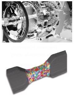
Image Credit: Carl Zeiss Microscopy GmbH
Ceramics
- Ceramics in 3D can be characterized
- Users can study grain growth in strontium titanate
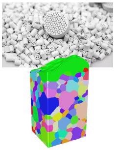
Image Credit: Carl Zeiss Microscopy GmbH
Semiconductors
- Semiconductor materials in 3D can be characterized
- Characterize huge grains and grain boundaries in polycrystalline silicon in affordable solar cells
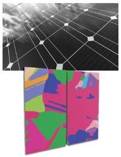
Image Credit: Carl Zeiss Microscopy GmbH
Pharmaceuticals
- Define pharmaceuticals in 3D
- Perform crystallographic mapping of hexamine crystals in 3D
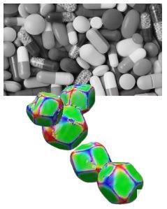
Image Credit: Carl Zeiss Microscopy GmbH
CrystalCT
Get Superior Sample Representivity from Advanced Diffraction Scan Modes
ZEISS Xradia Versa LabDCT Pro and Xradia CrystalCT advance materials characterization, modeling and discovery via innovative diffraction scanning modes.
- Offers unparalleled sample representivity
- Enhances speed
- Scanning of larger sample volumes
- Sample specificity has been fulfilled
- Streamlines sample preparation, and handling of uneven or realistic sample shapes
Inspired by nature’s golden angle, advanced scanning modes provide a helical phyllotaxis schema to regulate an extensive range of sample shapes and sizes and overcome a few of the early challenges of conventional DCT:
- Helical phyllotaxis is faster when the users’ sample is large and wide
- Helical phyllotaxis when users’ sample is tall and narrow
- Helical phyllotaxis is available with high aspect ratio tomography (HART) for flat samples
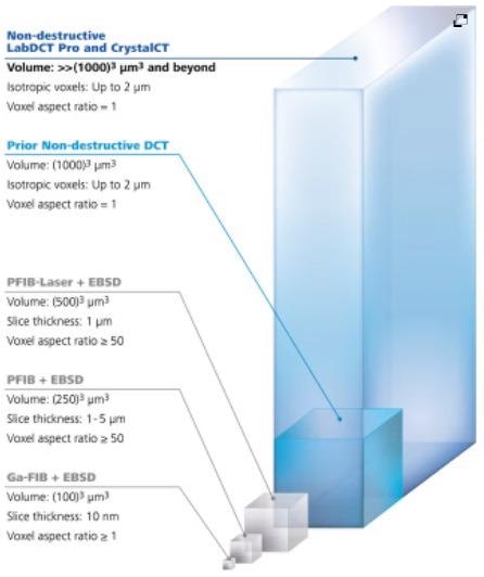
Image Credit: Carl Zeiss Microscopy GmbH
The Technology Behind LabDCT Pro and CrystalCT
DCT projection images tend to generate different diffraction patterns on the detector relying on their focusing geometries. As far as Laue focusing mode is concerned, grains produce diffraction spots consisting of sharp lines on the scintillator-coupled objective-based detector.
Projection geometry makes use of the flat panel detector placed in the magnifying or defocused position to gather diffraction spots that look like projected shape profiles of the equivalent diffracting grains.
The LabDCT Pro module on the ZEISS Xradia 620 Versa allows grain mapping on both the steadfast 4X DCT objective and on the optional flat panel detector offering the flexibility to select between high resolution or high throughput huge area grain mapping, whereas ZEISS Xradia CrystalCT utilizes its dedicated flat panel detector in projection mode.
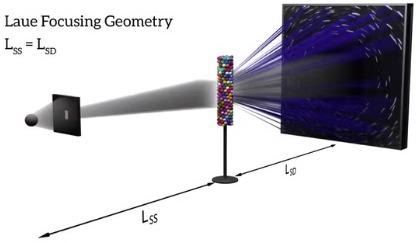
LabDCT Pro: Laue focusing geometry with the 4X DCT objective. Image Credit: Carl Zeiss Microscopy GmbH
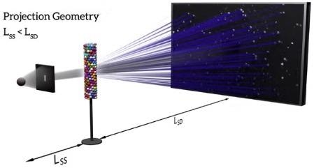
LabDCT Pro or CrystalCT: Projection or Laue focusing geometry onto a flat panel detector. Image Credit: Carl Zeiss Microscopy GmbH
3D Crystallographic Grain Mapping
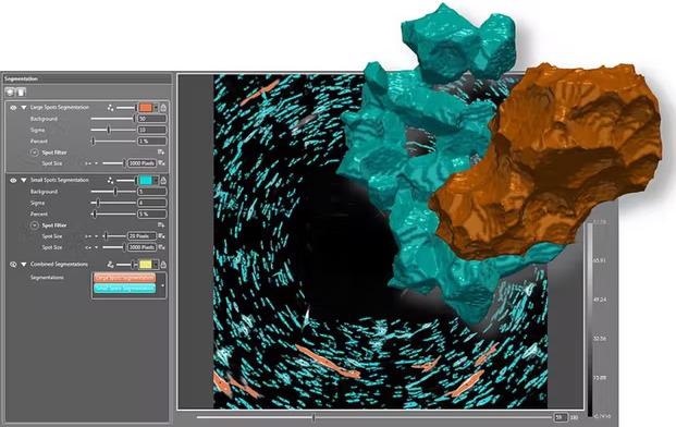
Image Credit: Carl Zeiss Microscopy GmbH
3D Grain Reconstruction
Index Grain Data Precise, Fast and Automated
Users can start to reconstruct after the initial acquisition as the first step in users’ workflow has been completed. Users can load their diffraction data and absorption tomography into GrainMapper3D. Let it determine possible candidates for grain orientations of a provided polycrystal with the help of back and forward projections.
The next step for the users is an automated, iterative search for grains in the sample volume. Grain reconstruction outcomes are stored as stacks of slices or volume datasets containing the complete description of the indexed grains. Ultimately, users can share 3D LabDCT Pro results with their collaborators or customers with the help of the independent GrainMapper3D Viewer application.
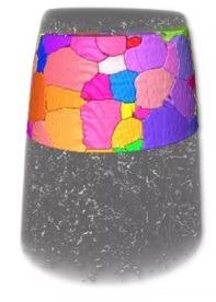
Image Credit: Carl Zeiss Microscopy GmbH
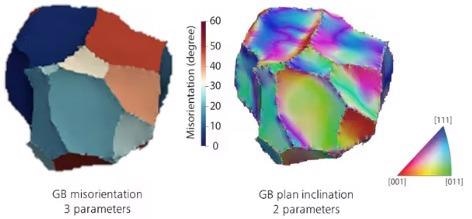
Image Credit: Carl Zeiss Microscopy GmbH
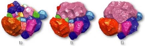
Image Credit: Carl Zeiss Microscopy GmbH
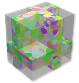
Image Credit: Carl Zeiss Microscopy GmbH
3D Grain Mapping
Get all Information in One File
The final step for users is to get all the data they need in a single file. The orientation, shape and spatial locations of all grains in the sample volume have been exported into an open data format.
Users can finish their experiments with consequent analyses by making use of simulation tools or customized software. The latest indexation routines currently support the more complicated lower symmetry crystal systems.
Novel Materials Research Enabled by Two Powerful X-Ray Solutions
ZEISS LabDCT Pro is considered to be an optional module for ZEISS Xradia 620 Versa, the highest performing and most versatile next generational X-Ray microscope with Resolution at a Distance (RaaD).
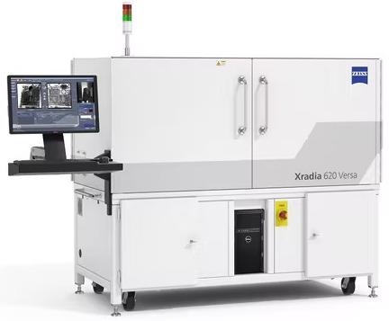
Image Credit: Carl Zeiss Microscopy GmbH
ZEISS Xradia CrystalCT isthe world’s first commercially available laboratory DCT built on a microCT platform, which is a highly-functional imaging system, thereby offering superb resolution and image quality for a range of 3D imaging requirements.
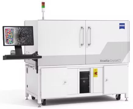
Image Credit: Carl Zeiss Microscopy GmbH