Users can organize and overlay photos from any source: Incorporate various dimensions and imaging modalities to get multiple perspectives on the sample. Using the ZEISS ZEN Connect software, users may combine all imaging techniques — ZEISS and non-ZEISS — to find answers to the queries.
- Utilize the overview image for navigation
- Obtain an overview image
- Save all multi-modal data in well-organized projects
- Transfer to the light, confocal or electron microscope and align once
- Easily work with ZEN Connect to line up and overlay images from any source
ZEISS ZEN Connect Always Shows Your Data in Context
Whether the user is a materials researcher at a university, a multi-use facility or an industrial lab, he or she may obtain new insights, increase efficiency and save time.
When users want to characterize the qualities of your sample in-depth, they often need to use a combination of microscopy modalities or contrasting approaches.
Whether the user is looking for e-mobility batteries or experimenting with additive-produced parts, ZEN Connect is there to rescue. Oil and other basic resources may pique curiosity. Alternatively, determine whether metallic inclusions in a material sample exist. A single contrast or microscopy method will not be enough to address all of the queries in these and other situations.
Users can benefit from having more than one microscopy method at their disposal.
Highlights
Overlay and Align Images
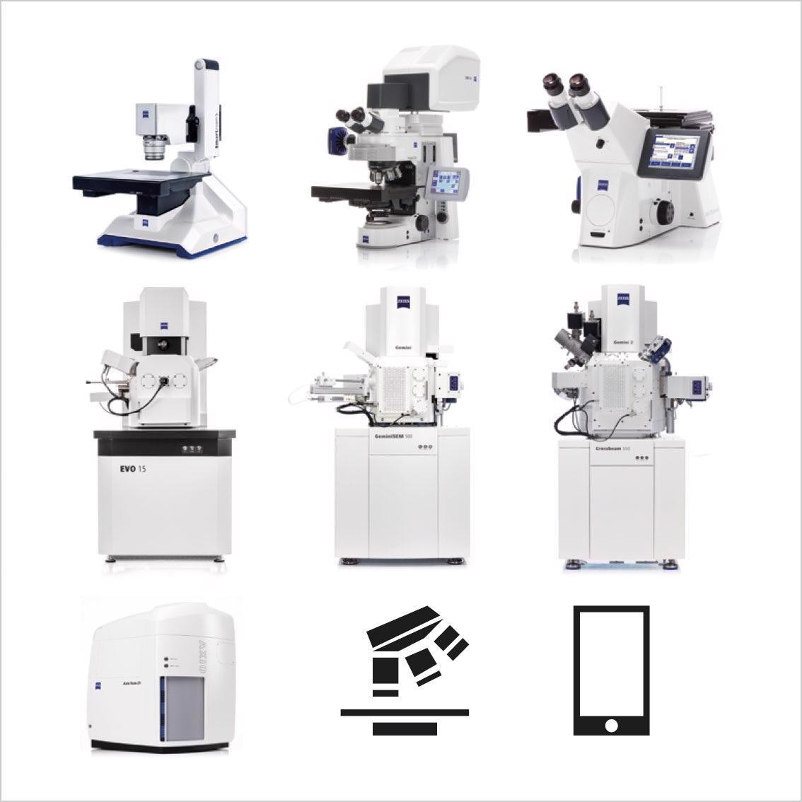
Image Credit: Carl Zeiss Microscopy GmbH
- Load complex multi-dimensional images or just simple overview images from users’ mobile phone
- It makes no difference whether the imaging technology is from ZEISS or from a third party
- All image data can be associated and shown in context
- ZEISS ZEN Connect keeps its metadata so long as external images track to the well-established Bio-Formats standard
Acquire Overview Images for Easy Navigation
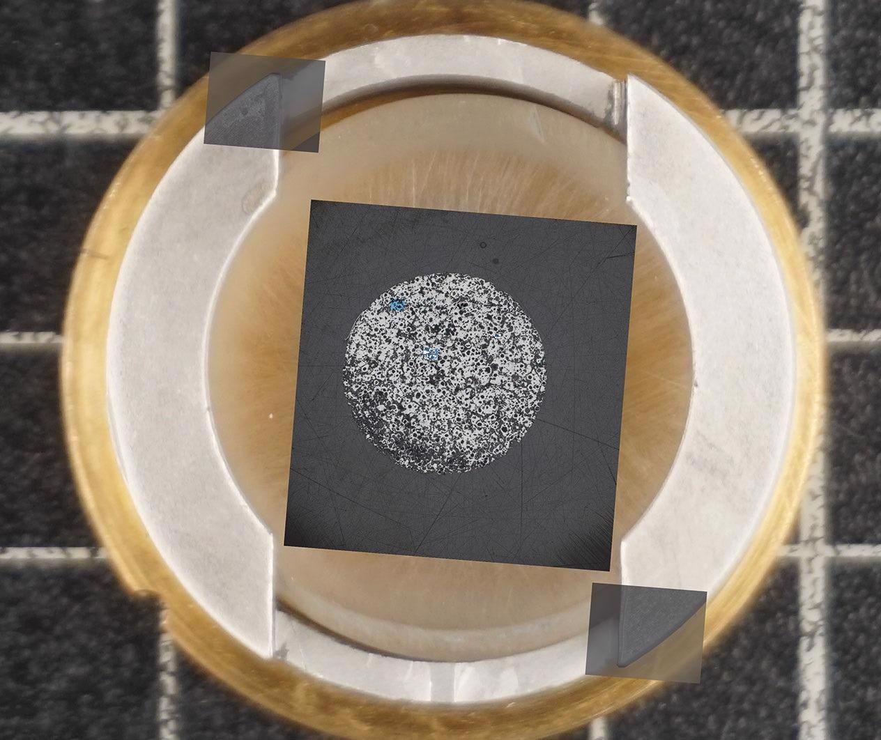
Image Credit: Carl Zeiss Microscopy GmbH
- Discover all subsequent images in context as users zoom in and out across the borders of resolution domains and imaging technologies
- Image the sample with a ZEISS stereo microscope or any other low magnification system
- Mount the specimen on a sample holder from ZEISS or any other independent one
- Take the stage to the right position with a simple click on the overview image and examine or re-evaluate your ROIs.
- Utilize the overview image to navigate and find the ROIs
Smart Data Management
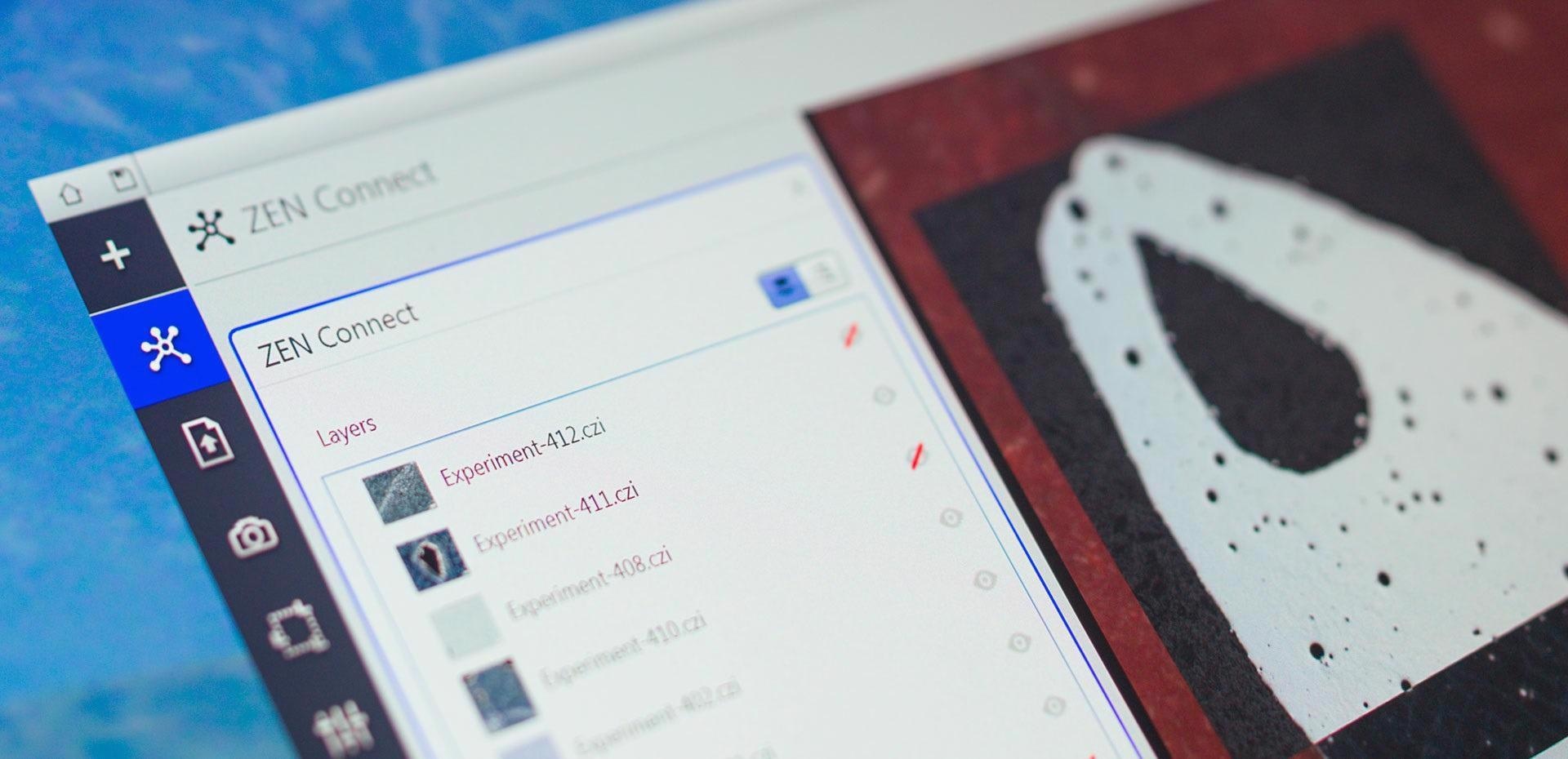
Image Credit: Carl Zeiss Microscopy GmbH
- All images are protected in database projects with an intuitive label attached automatically
- Stay on top of things — during the experiments or in the months afterward
- Discover all the images and their connected datasets
- Examine for microscope type and imaging parameters with the filter function
Applications
Materials Research in E-Mobility
Mastering permanent magnets are critical to the future of e-mobility. Begin by obtaining an overview photograph. Kerr microscopy can be used to see magnetic domains. Determine which magnetic phases are the most promising. From the micro to the nanoscale scale, reveal features and boost resolution.
Draw inferences regarding the sample morphology and magnetic properties’ relationship. Connect broad field-of-view photographs with the best possible resolution at all times during the experiment to analyze the data in a larger context.
Large-scale overview, acquired with ZEISS Axio Imager. Image Credit: T. Schubert, Aalen University, Germany
Magnetic domain structure visualized by Kerr microscopy. Image Credit: T. Schubert, Aalen University, Germany
Correlated high-resolution image acquired with ZEISS Sigma 300 VP. Image Credit: T. Schubert, Aalen University, Germany
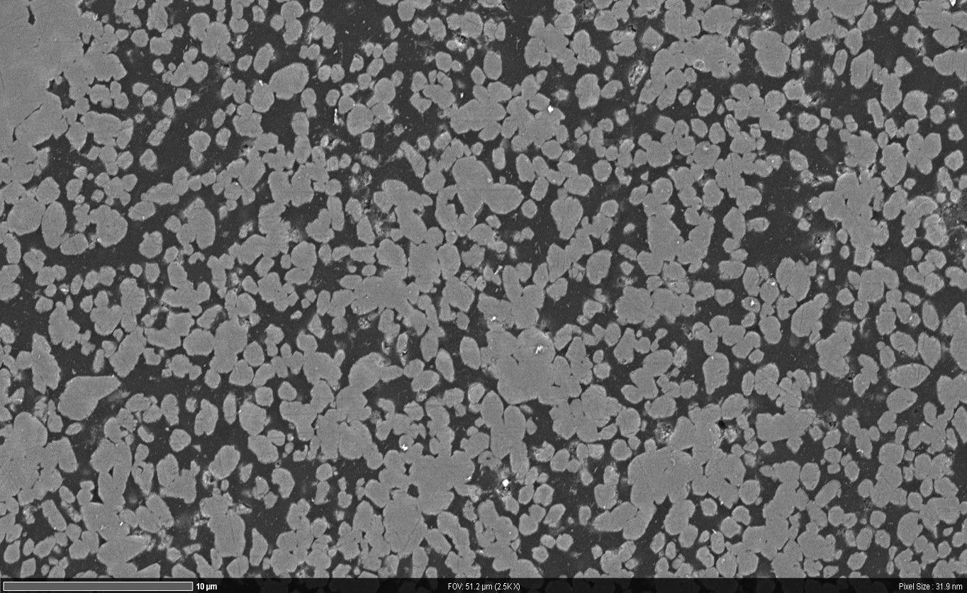
Materials Research on Inclusions in Calcium-treated Steel
In steel, calcium treatment prevents the formation of harmful manganese sulfide (Mn) inclusions. These inclusions can extend while rolling, causing steel anisotropy. When Mn inclusions are converted to the much harder calcium sulfide (CaS) inclusions, they remain globular and contribute to the steel’s isotropy. Combine photos into a single large-scale overview image of the entire sample.
Rapidly identify and screen huge numbers of inclusions. Following that, users may want to go from overview to detail. For high-resolution imaging, switch to SEM. Select specific inclusions for EDS analyses by relocating the region of interest.
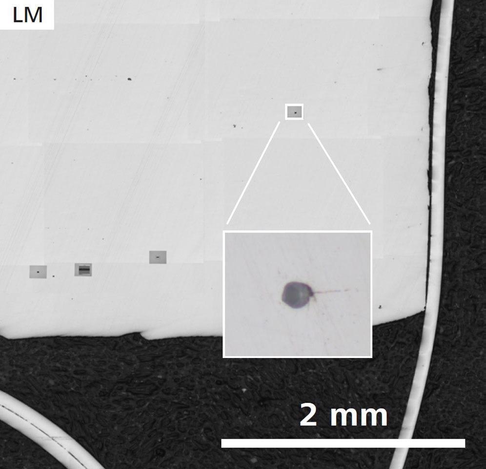
First sample, without Ca treatment: Sample Overview and detail of one inclusion (inset) acquired by ZEISS Axio Observer. Image Credit: T. Schubert, Aalen University, Germany
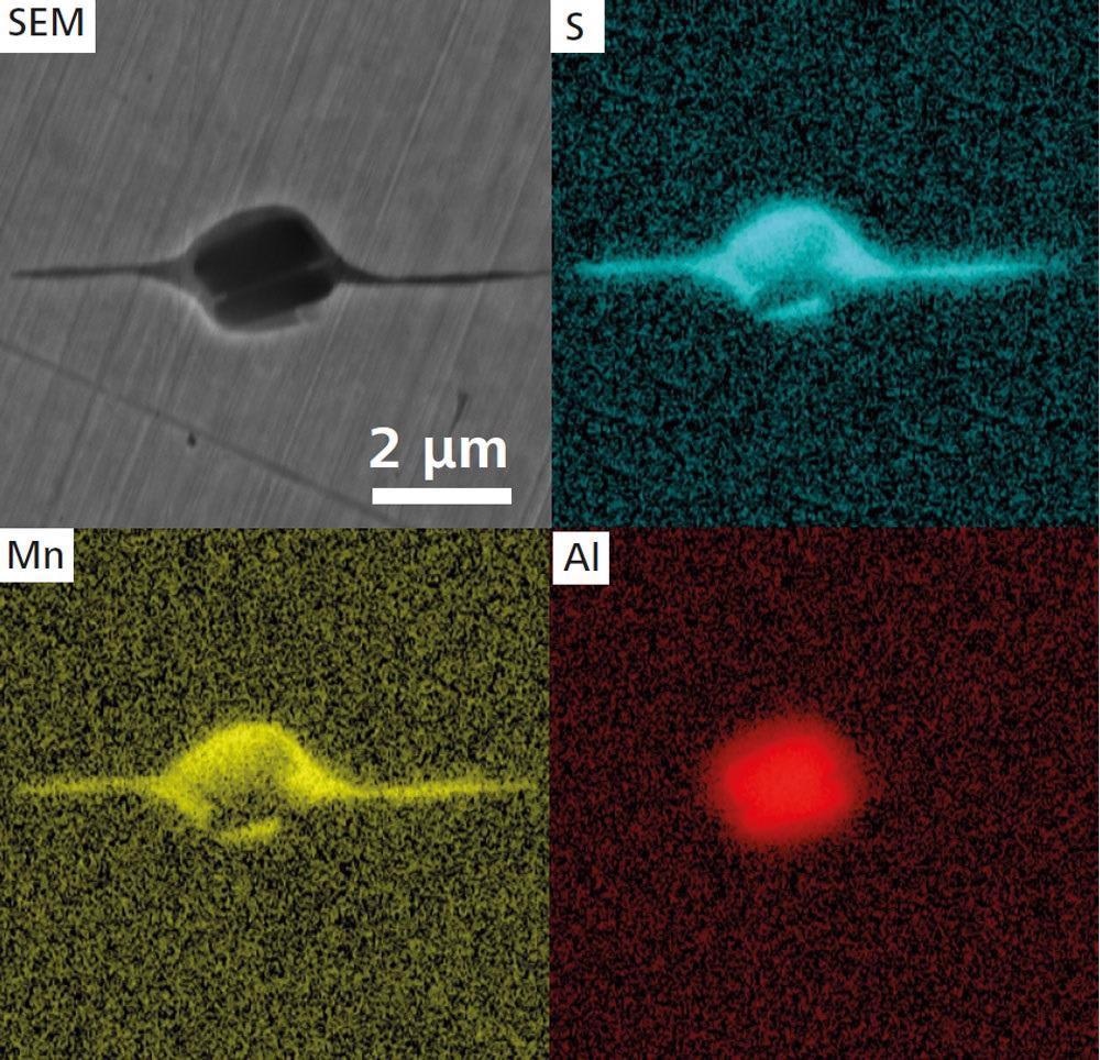
Higher magnification SEM image (ZEISS Crossbeam 550) and EDS maps of an elongated MnS inclusion. Image Credit: T. Schubert, Aalen University, Germany
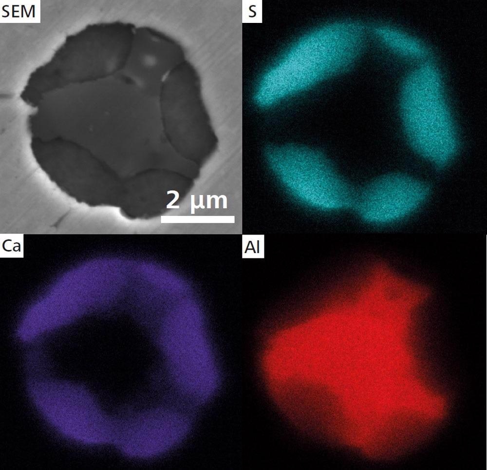
Second sample with Ca treatment SEM image of a globular CaS inclusion and EDS maps. Image Credit: T. Schubert, Aalen University, Germany
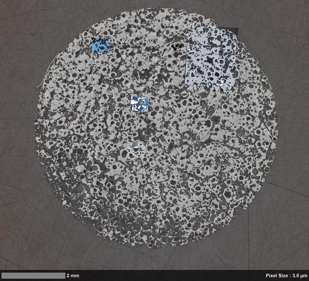
Materials Research in Oil Recovery, Nuclear Waste Disposal and Carbon Capture and Storage
Carbonate rocks have pore structures that users should be aware of. Characterize flow, response and transport over a wide variety of length scales and in the presence of significant structural variation. Gain access to critical data during the recovery of oil, carbon capture and storage and the disposal of nuclear waste. Automated light microscopy is used to image a broad area.
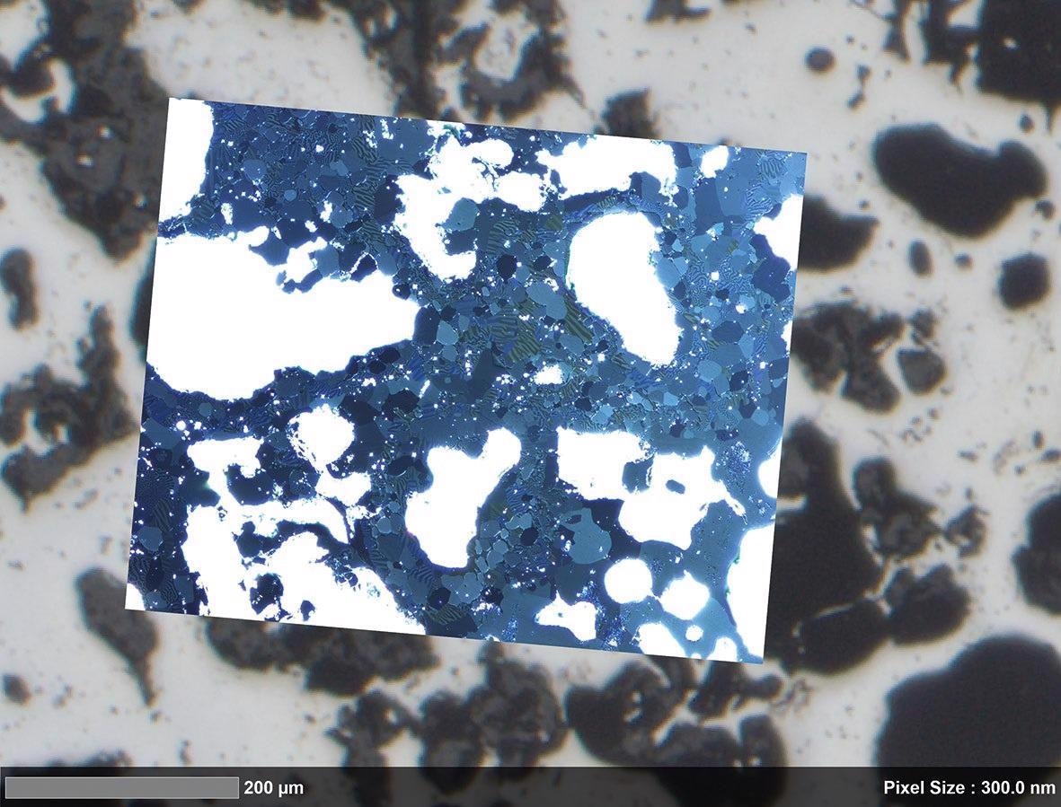
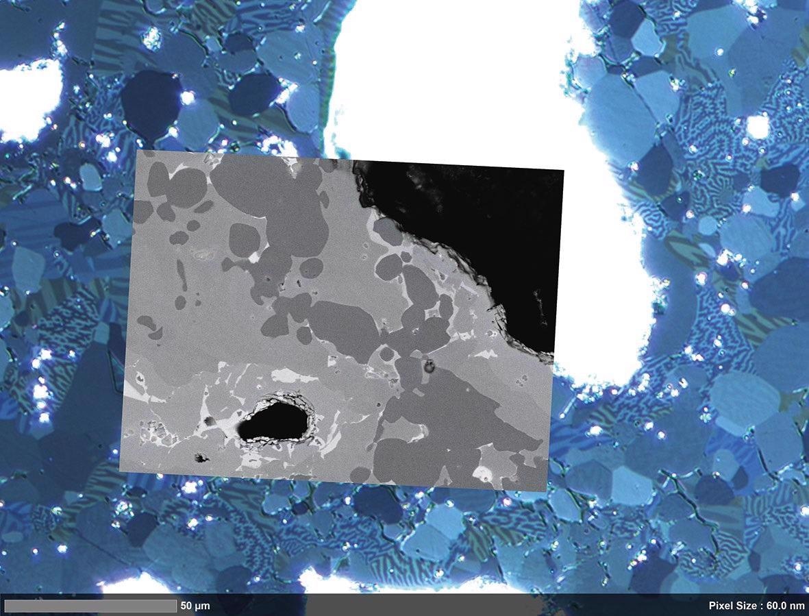
Import the image into ZEISS ZEN Intellesis for complete macro-structure classification, identifying macro-pores, solid rock grains and microporous grains based on color, texture and intensity changes. Use this information to find areas of interest for high-resolution field emission scanning electron microscopy. When studied in the context of macroscopic heterogeneity, users can notice changes in the local nanometer-scale pore structure.
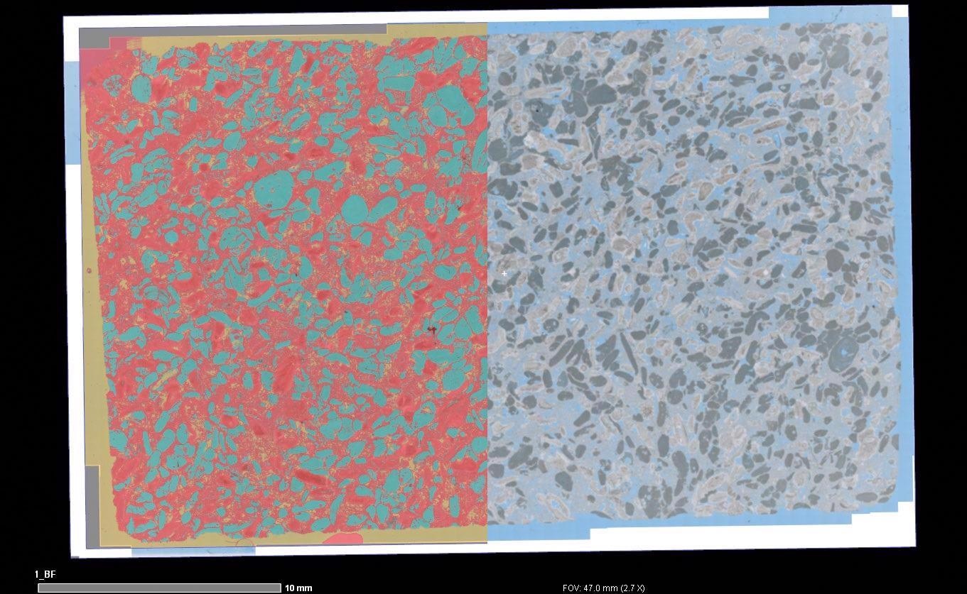
Overlay of macroscopic segmentation (left) and automated light microscopy image (right) showing the microporosity and microporous grains. Image Credit: T. Schubert, Aalen University, Germany
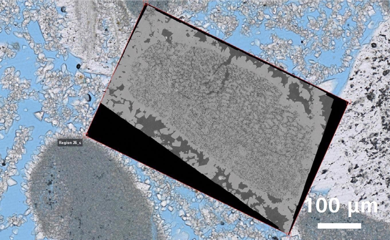
Correlative overlay of high-resolution light microscopy data and nano-scale electron microscopy data. Image Credit: T. Schubert, Aalen University, Germany
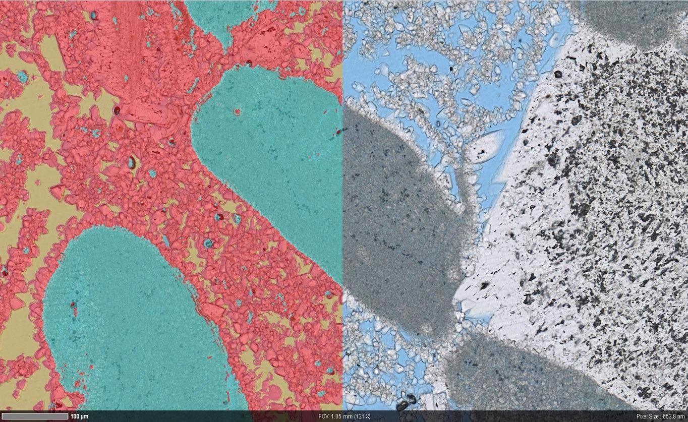
Overlay of segmented (left) high resolution light microscopy data (right) showing the pore structure in more detail. Image Credit: T. Schubert, Aalen University, Germany
High resolution nano-scale microscope data. Image Credit: T. Schubert, Aalen University, Germany
Accessories
ZEISS ZEN Data Storage
Central Data Management in the Connected Laboratory
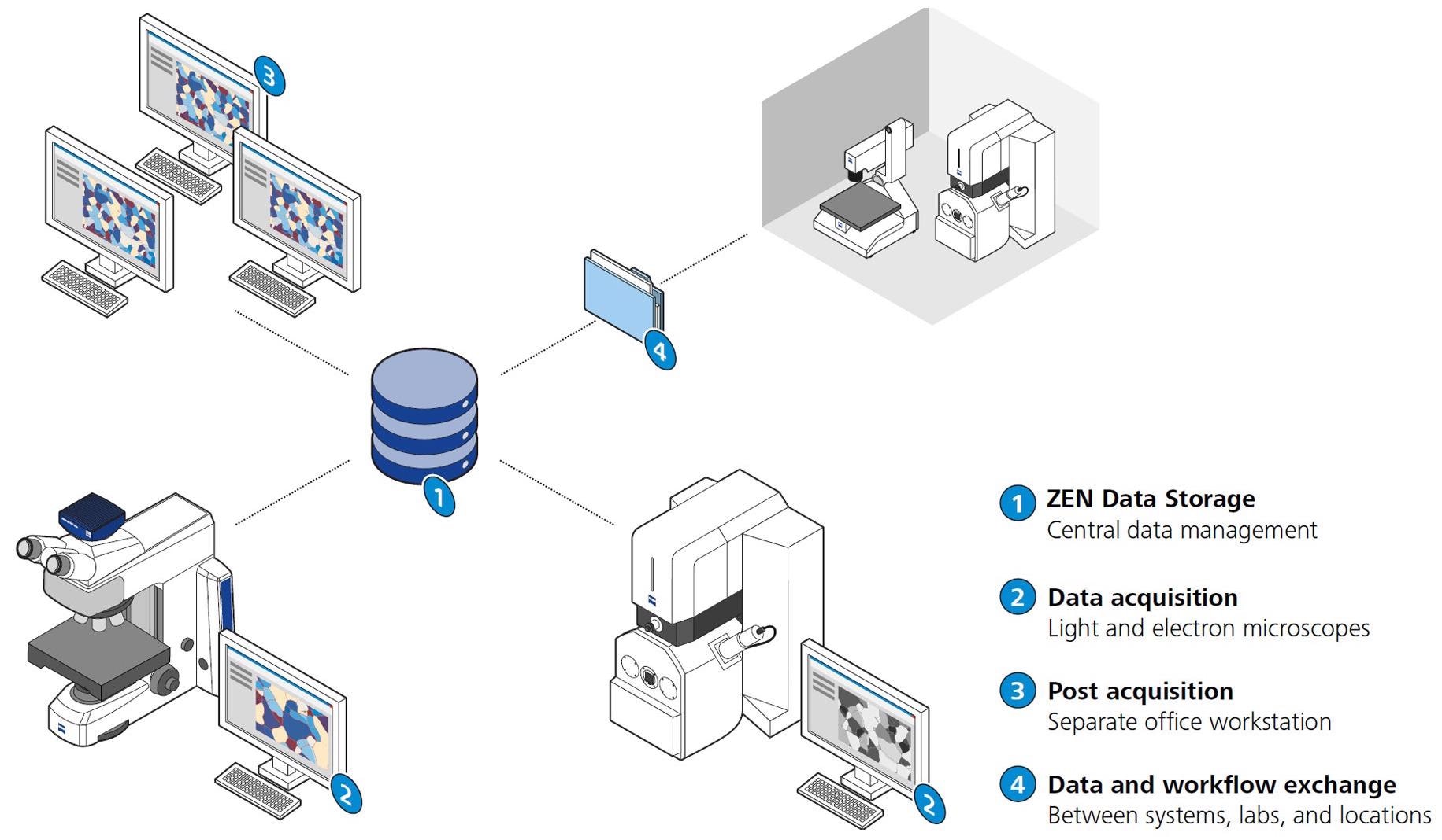
Image Credit: Carl Zeiss Microscopy GmbH
As digitization improves microscopic investigations, users will be confronted with an ever-increasing number of images and data to manage, especially in multi-user laboratories. Image and data capture may be separated from post-acquisition work using ZEISS ZEN Data Storage, allowing everyone in the lab to work more efficiently. Access to data and sharing capabilities assist the collaborators as well.
Correlative Microscopy with ZEISS Shuttle & Find
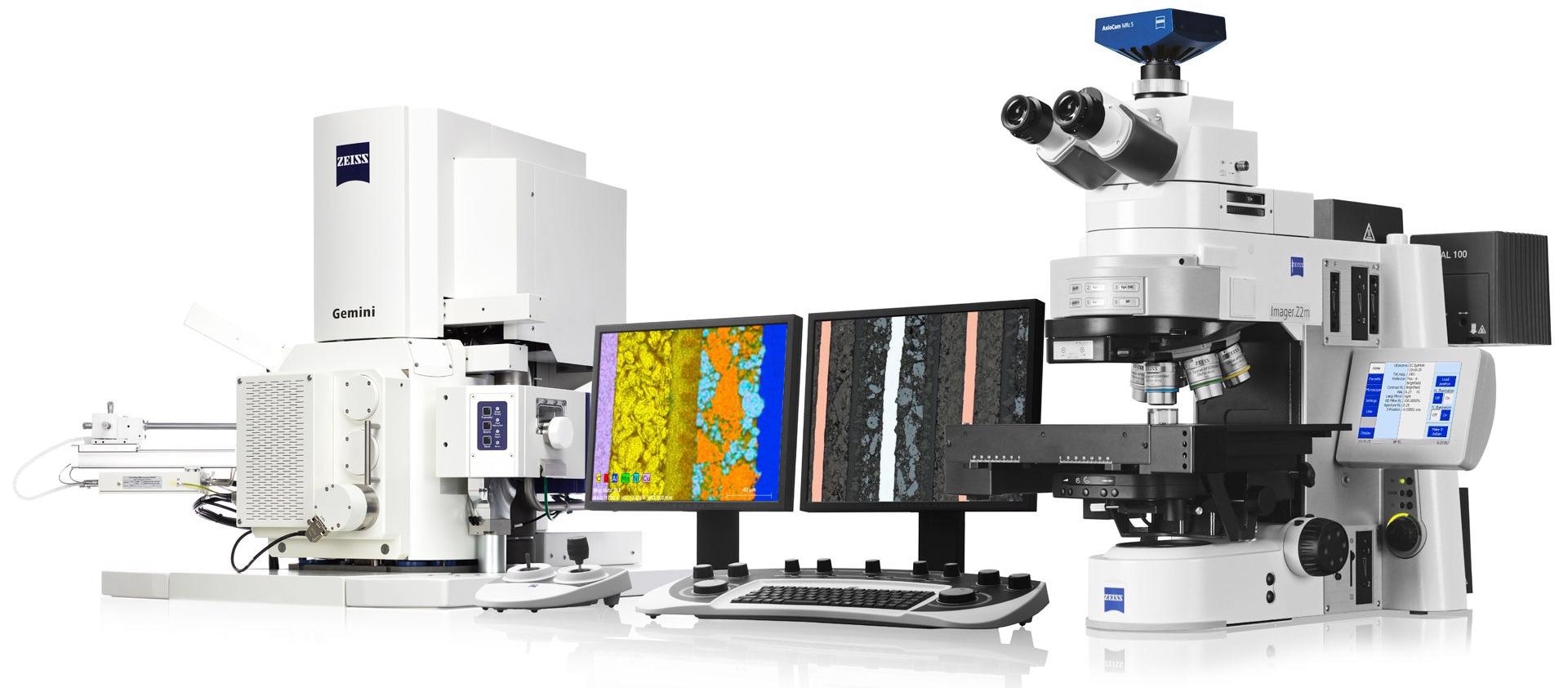
Image Credit: Carl Zeiss Microscopy GmbH
With the ZEISS Shuttle & Find software module, users can overlay data from their light microscope and scanning electron microscope in a convenient, efficient workflow. To create a coordinate system in seconds, they can use a specimen holder with fiducial markers.
Using a light microscope, define regions of interest in the sample. Get access to the highest imaging and analytics by relocating the ROIs in the electron microscope. Lastly, compare and contrast the images obtained using various microscopical techniques.
ZEISS ZEN Intellesis for Image Segmentation in Microscopy

Image Credit: Carl Zeiss Microscopy GmbH
Use deep learning to quickly separate your photographs and gain access to their true value—the data they contain. All subsequent image analysis procedures are built on the foundation of picture segmentation.
Deep learning and Python are used by ZEISS ZEN Intellesis to simply provide repeatable segmentation results, even for non-experts. After training the software, ZEISS ZEN Intellesis can automatically segment a batch of hundreds of photos. Users can save time while reducing user bias.